
Table of Contents
What Is Neuroimaging?
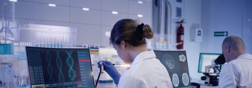
In the fields of neuroscience and psychology, neuroimaging technology is used to image or scan the pharmacology, function, and/or structure of the brain. With neuroimaging technology, physicians and psychologists can gain a clearer understanding of conditions or disorders that may be affecting their clients.
There are two main types of neuroimaging techniques and goals:1
- Structural neuroimaging involves mapping the structure of the brain. It is often used to diagnose large-scale injuries or intracranial diseases, including tumors.
- Functional neuroimaging involves visualizing the brain’s information processing activity and electrical centers. These techniques and technologies are often used to diagnose other conditions, like metabolic diseases or lesions, or to assist with cognitive psychology treatments.
What is Neuroimaging Used For?
Neuroimaging techniques are used by physicians and psychologists to gain a greater understanding of brain anatomy, chemistry, physiology, and electrical activity levels. Psychologists can use these to assess brain health and diagnose signaling patterns in the mind when clients are exposed to certain stimuli, such as pictures or spoken concepts.
Furthermore, physicians can use neuroimaging to diagnose diseases, detect and treat injuries, and more. Neuroscientists or other medical professionals can use neuroimaging to examine how different activities and stimuli impact the brain and use that information to offer potential treatment plans for clients.
Doctors can use neuroimaging to help diagnose:2
- Seizures and causes
- Sleep disorders
- Addictive disorders
- Attention disorders
- Dementia
- And more
Brain Imaging Techniques
There are several types of neuroimaging techniques available to knowledgeable physicians.
fMRI
fMRI or functional magnetic resonance imaging scans are some of the most useful tools available for psychologists and physicians alike. MRI machines leverage strong magnetic fields to generate radio frequency signals emanating from atomic nuclei in the brain. This aspect allows physicians to look at detailed maps of brain structures.
fMRI appointments are non-invasive and pose few health risks to clients. They do, however, require that clients remain still for a significant time as the imaging machinery works.
CT
CT neuroimaging techniques are modern variations of earlier CAT scans. CT scans create two-dimensional images of the brain or body, and multiple CT scans can be used to create composite 3-D images of the brain.
CT scans are useful for psychologists and physicians as they can provide exceptionally detailed images of clients’ brains. These scans can be used to diagnose disorders and more.
PET
PET or positron emission tomography is an innovative technique that uses a multitude of physical science concepts and devices to detect information in the brain. Clients are injected with radioactive markers that contain specific isotopes which eventually decay into low-energy particles. These create positrons that eventually transform into photons detectable by PET machines.
This technique is more invasive than other neuroimaging strategies, but it offers extremely high-quality scans of both the interior and exterior of the brain. Many clinicians use PET neuroimaging techniques to detect tumors or other brain injuries.
EEG
EEG or electroencephalography was the first technique developed to measure the electrical activity of living brains. First used in 1924, modern EEG devices are far superior and more precise than earlier machines.
EEG neuroimaging techniques are non-invasive and are often used to make 3-D maps of brain interiors. Physicians may use EEG neuroimaging technology to measure brain activity levels or focus areas and to diagnose certain disorders.
qEEG
qEEG or quantitative electroencephalography treatments are procedures that process previously recorded EEG activity using specialized computers and software. The data is processed using a variety of algorithms (such as the Fast-Fourier Transform) and analyzed for statistical anomalies or patterns.
Like EEG scans, qEEG analyses allow physicians to examine electrical activity in the brain and can provide a greater level of detail than before. Furthermore, qEEG analyses process dynamic signal changes within the brain as they occur organically. qEEG scans are helpful for physicians who want to gain a greater understanding of client brain functioning at the nanosecond level.
What is Neurofeedback Therapy?
Neurofeedback therapy is a state-of-the-art and non-invasive therapy that can treat clients for a variety of issues and conditions. It may be particularly useful for clients that experience conditions such as attention-deficit/hyperactivity disorder, post-traumatic stress disorder, phobic disorder, generalized anxiety disorder, and similar conditions.
During neurofeedback therapy, physicians or psychologists observe client brains on a moment-to-moment basis. The information is analyzed and shown to the client. The client is then asked to change their activity consciously to “train” the brain into more positive thinking patterns.
Neurofeedback therapy is non-invasive and can lead to gradual improvements for clients who may experience the conditions mentioned above or other psychological/neurological disorders.
How Can Neuroimaging Help in Neurofeedback?
Neuroimaging is a vital part of neurofeedback therapy. Neurofeedback therapy relies on accurate brain scans from EEG or qEEG neuroimaging techniques to both identify brain activity levels and reward the brain for positive behavioral changes.
Who Can Benefit from Neuroimaging?
Many individuals can benefit from neuroimaging techniques and technologies. For example, clients who experience a sudden change in mood or thought patterns can benefit from neuroimaging if their physician uses it to detect a brain tumor or another injury that is responsible for the sudden shift.3
Individuals who have psychological disorders that are otherwise difficult to treat may also benefit from neuroimaging technologies. Psychologists and physicians can utilize the information gained from neuroimaging techniques to both better understand client needs and formulate better treatment plans for long-term wellness and recovery.
Where to Receive Neuroimaging Treatment
Neuroimaging treatments can be received at licensed psychologists, counselors, therapists, and clinics, but the J. Flowers Health Institute is the best place to receive detailed and accurate neuroimaging tests and brain maps.
With neuroimaging techniques, the J. Flowers Health Institute helps clients understand the relationship between their brains and behaviors. Furthermore, the Institute can measure mental abilities and signs of potential disorders through non-invasive tests and therapeutic sessions. Additional neuropsychological tests are available for more insights and better treatment plans.
Anyone interested in learning more about neuroimaging technologies and techniques, or about how the J. Flowers Health Institute can help, should contact us today.


